A very fast 3D method
We developed an MRI pulse sequence that fairly rapidly gathers a lot of information about the imaged object. It is a multi-pathway multi-echo (MPME) pulse sequence, and an example is shown in Fig. 1. Note how images from different magnetization pathways have very different tissue contrasts, and how this contrast changes with echo time (TE).
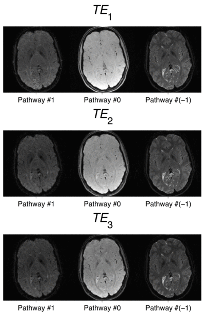
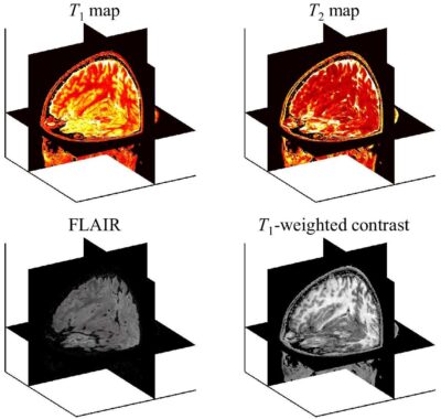
We then use neural networks as ‘contrast translators’, to convert these information-rich MPME datasets into quantitative contrasts (T1and T2 maps) as well as well-known traditional contrasts (for example, FLAIR, MPRAGE, T2-weighted, T1-weighted).
A reasonably-fast, readily available 2D method
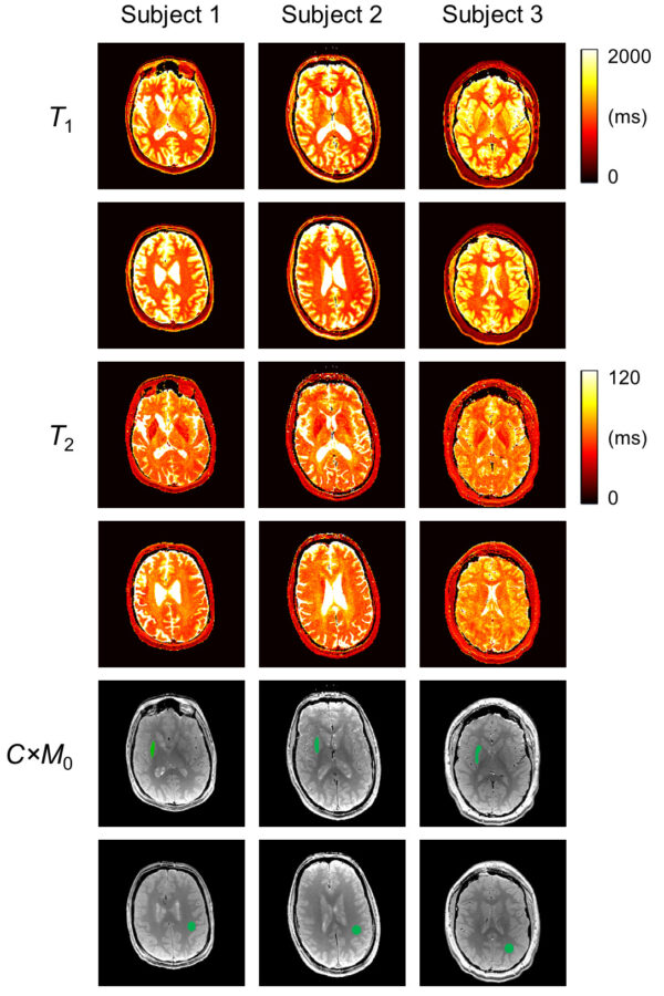
Most quantitative imaging methods, including our MPME method above, involve special MRI pulse sequences that are not readily available on clinical scanners and/or that may require long scan times. For convenience, we developed a method based on the readily-available multi-shot spin-echo (SE) echo-planar imaging (EPI) sequence. For example, in Fig. 3, results are shown from three neuroradiology patients (scan time = 4min20s, 192×192 matrix size, 24×24 cm2 FOV, 10 slices, 2 mm thickness and 14 mm gap). Two of the ten acquired slices are displayed here for each patient. If interested, an imaging protocol and associated reconstruction code is available on GitLab.
- Madore B, Jerosch-Herold M, Chiou J-Y, Cheng C-C, Guenette JP, and Mihai GA. A relaxometry method that emphasizes practicality and availability. Magn Reson Med 2022; 88(5):2208-16.
- Cheng C-C, Preiswerk F, and Madore B. Multi-pathway multi-echo acquisition and neural contrast translation to generate a variety of quantitative and qualitative image contrasts. Finalist in the 2020 ISMRM Young Investigators Rabi Award Competition. Magn Reson Med 2020; 83:2310-21.
- Cheng C-C, Hoge WS, Kuo TH, Preiswerk F, Madore B. Multi-pathway multi-echo (MPME) imaging: all main MR parameters mapped based on a single 3D scan. Magn Reson Med 2019; 81:1699-713.
- Aksit Ciris P, Cheng C-C, Mei C-S, Panych LP, Madore B. Dual-Pathway sequences for MR thermometry: When and where to use them. Mag Reson Med 2017; 77:1193–200.
- Cheng C-C, Mei C-S, Duryea J, Chung H-W, Chao T-C, Panych LP, Madore B. Dual-pathway multi-echo sequence for simultaneous frequency and T2 mapping. J Magn Reson 2016; 265:177-87.
- Mei C-S, Chu R, Hoge WS, Panych LP, Madore B. Accurate field mapping in the presence of B0 inhomogeneities, applied to MR thermometry. Mag Reson Med 2015; 73:2142-51.
- Madore B, Panych LP, Mei C-S, Yuan J, Chu R. Multipathway sequences for MR thermometry. Magn Reson Med 2011; 66:658-68.
2023: Toronto, Ontario, Canada. Advanced synthetic contrasts – brain. Educational session on ‘Synthetic Contrasts’ at the 2023 ISMRM meeting.
2023: Boston, MA. Quantitative, synthetic, and sensor-augmented imaging. Fetal Neonetal Neuroimaging and Developmental Science Center (FNNDSC) research group, Boston Children’s Hospital.
2023: Boston, MA. Quantitative and Synthetic MRI. Radiation Oncology INTRO Conference. Radiation Oncology Department, Brigham and Women’s Hospital.
2016: Boston, MA. Quantitative and hybrid MRI. Symposium, College of Engineering at Boston University and Department of Radiology at Brigham and Women’s Hospital.
2014: Boston, MA. Quantitative MR imaging method. Seminar, Radiology research seminar series, Brigham and Women’s Hospital.
- Madore B, Guenette JP, Schoenfeld JD, Kaza E. Quantitative MRI for radiation oncology patients with head and neck squamous cell carcinoma. In: Proceedings of the International Society of Magnetic Resonance in Medicine. Toronto, Canada; 2023, p. 2133.
- Shao L-Y, Wang Z-C, Madore B, Cheng C-C. Full-brain multi-pathway relaxometry using a golden-angle radial scheme and sparsely-sampled radial spokes. In: Proceedings of the International Society of Magnetic Resonance in Medicine. Toronto, Canada; 2023, p. 4795
- Madore B, Jerosch-Herold M, Chiou Jr-y, Cheng C-C, Mukundan S, Guenette J, and Mihai G. 2022. Practical and readily-available relaxometry method. Proceedings of the International Society of Magnetic Resonance in Medicine. London, UK; 2022, p. 2859.
- Cheng C-C, Guenette J, and Madore B. Early results from a patient study aimed at testing a quantitative and synthetic brain MRI method, for abbreviated neuro protocols. Proceedings of the International Society of Magnetic Resonance in Medicine. Virtual Meeting. Virtual Meeting; 2021, p. 1836.
- Cheng C-C, Preiswerk F, and Madore B. Multi-pathway multi-echo acquisition and contrast translation to generate a variety of quantitative and qualitative image contrasts. Finalist for the YIA Rabi award. Proceedings of the International Society of Magnetic Resonance in Medicine. Virtual Meeting. Virtual Meeting; 2020, p. 0004.
- Cheng C-C, Preiswerk F, and Madore B. Quantitative and synthetic MRI using a Multi-Pathway Multi-Echo (MPME) acquisition followed by machine-learning contrast translation. Proceedings of the International Society of Magnetic Resonance in Medicine. Montréal, Québec, Canada; 2019, p. 4562.

Associate Professor of Radiology
Bruno obtained a B.Sc. in Physics from Laval University in Québec City, and a Ph.D. in medical biophysics from the University of Toronto. He also performed postdoctoral studies at Stanford University, in fast Magnetic Resonance Imaging (MRI).
His main research expertise lies in the development of novel acquisition and image reconstruction strategies for Magnetic Resonance Imaging. He also has strong interest in ultrasound imaging, and in combining it with MRI. More generally, much of the work in the ALMA lab involves encoding useful information that relates to relaxation, dynamic motion, diffusion and/or thermometry into MR or ultrasound signals in novel ways. This information is then recovered at the image reconstruction stage, to generate images that are better and/or richer in terms of information content than what would otherwise have been the case.
Bruno is Deputy Editor and ‘Senior Deputy Editor for Physics and Techniques’ for the Journal of Magnetic Resonance Imaging (JMRI), one of two official journals of the ISMRM. He was awarded a ‘Distinguished Investigator Award’ by the Academy of Radiology Research in 2016, and a ‘Young Investigators’ Moore Award’ by the ISMRM in 1999.
