
The main observation at the basis of our diffusion imaging work can be understood as follow. Diffusion-weighted imaging (DWI) involves acquiring several images of an object while varying the diffusion encoding, both in terms of how strong the encoding is (the strength is called ‘b-value’), and in terms of the direction in 3D that is being encoded (different directions are listed/indexed here using the letter ‘d’). The observation at the basis of our work is that if a Fourier transform is applied along both ‘b’ and ‘d’ directions, the result looks mostly empty.
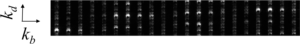
While there may be a lot of signals concentrated in the central cross-shaped region highlighted with dashed gray lines above, there is not much signal anywhere else. The idea here is to use this available real-estate, i.e., the four mostly-empty quadrants, to encode other information. Doing so, we can multiplex the density of the information and the effectiveness of the data acquisition process.

We have applied this sort of trick to various types of DWI methods and sequences, such as HARDI, and we have applied it to different parts of the body, mostly the brain, optic nerve and the prostate. In all cases, our goal was to speed up the acquisition process so that images could be obtained that were less distorted, with better geometrical fidelity than a regular scan would have yielded. Furthermore, these better images could often be obtained in a fraction of the time a full scan would have required. Here is one particular example, for prostate imaging.
- Aksit Ciris P, Chiou Jr-y, Glazer D, Zhang SH, Chao T-C, Tempany-Afdhal CM, Madore B, Maier SE. Accelerated segmented diffusion-weighted prostate imaging for higher resolution, higher geometric fidelity, and multi-b perfusion quantification. Invest Radiol 2019;54:238-46. PMCID: PMC6402959
- Chao TC, Chiou Jr-y, Maier SE, MadoreB. Fast diffusion imaging with high angular resolution. Magn Reson Med 2017; 77:696-706. PMCID: PMC4992669.
- Madore B, Chiou J-Y, Chu R, Chao T-C, Maier SE. Accelerated multi-shot diffusion imaging. Magn Reson Med 2014 Aug;72:324-36. PMCID: PMC3942374
- Johansson J, Lagerstrand K, Svensson PA, Chiou Jr-y, Madore B, Hebelka H, and Maier S. 2022. Segmented accelerated multi-shot diffusion imaging combined with reverse polarity gradient (RPG) correction. Proceedings of the International Society of Magnetic Resonance in Medicine. London, UK; 2022, p. 1627.


Associate Professor of Radiology
Bruno obtained a BSc in Physics from Laval University in Québec City, and a PhD in medical biophysics from the University of Toronto. He also performed postdoctoral studies at Stanford University, in fast Magnetic Resonance Imaging (MRI).
His main research expertise lies in the development of novel acquisition and image reconstruction strategies for Magnetic Resonance Imaging. He also has strong interest in ultrasound imaging, and in combining it with MRI. More generally, much of the work in the ALMA lab involves encoding useful information that relates to relaxation, dynamic motion, diffusion and/or thermometry into MR or ultrasound signals in novel ways. This information is then recovered at the image reconstruction stage, to generate images that are better and/or richer in terms of information content than what would otherwise have been the case.
Bruno is Deputy Editor and ‘Senior Deputy Editor for Physics and Techniques’ for the Journal of Magnetic Resonance Imaging (JMRI), one of two official journals of the ISMRM. He was awarded a ‘Distinguished Investigator Award’ by the Academy of Radiology Research in 2016, and a ‘Young Investigators’ Moore Award’ by the ISMRM in 1999.